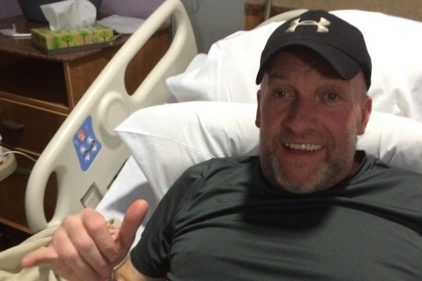
“It turned out that it was not benign, but it was a malignant glioblastoma, a very very aggressive cancer,” he explained.Īngela DeCellis and her husband, Matt, and their friend Andrea Hoffman. Doctors thought it was benign at first, but the pathology results indicated otherwise, Al Lupiano recalled. A scan revealed that she had a tumor on the left side of the brain that was 30 millimeters. “I was highly motivated to keep moving forward.” Brain tumor diagnosis within weeksĭeCillis started showing stroke-like symptoms and went to the hospital in August 2021. You have to find out if it’s something that’s going to affect my kids,’” Al Lupiano, 50, of Jamesburg, New Jersey, told TODAY. “We had talked about it from day one, that there’s something wrong here, and because of my sister’s background in medicine, she had said the same thing: ‘You need to figure this out. As DeCillis’ health declined, Al Lupiano started wondering why the three of them had brain tumors.

Last year, his wife, Michele Lupiano, was diagnosed with an acoustic neuroma and his sister, Angela DeCillis, was diagnosed with a glioblastoma, an aggressive brain tumor. He was successfully treated for it, though he has lingering side effects, including slight deafness, fatigue and dizziness. All rights reserved.In 1999, Al Lupiano says he was diagnosed with an acoustic neuroma, a type of benign brain tumor that grows on a nerve that runs from the brain to the inner ear. Glioma Inverse modeling MRI Model calibration Physics-based deep learning Tumor modeling.Ĭopyright © 2022 Elsevier B.V. We believe the proposed inverse solution approach not only bridges the way for clinical translation of brain tumor personalization but can also be adopted to other scientific and engineering domains. Coined as Learn-Morph-Infer, the method achieves real-time performance in the order of minutes on widely available hardware and the compute time is stable across tumor models of different complexity, such as reaction-diffusion and reaction-advection-diffusion models. In this work, we introduce a deep learning based methodology for inferring the patient-specific spatial distribution of brain tumors from T1Gd and FLAIR MRI medical scans. However, the unifying drawback of all existing approaches is the time complexity of the model personalization which prohibits a potential integration of the modeling into clinical settings. Alongside, various parametric inference schemes were developed to perform an efficient tumor model personalization, i.e. It includes different mathematical formalisms describing the forward tumor growth model. Over recent years a corpus of literature on medical image-based tumor modeling was published.
Kevin renderman brain tumor full#
To estimate tumor cell densities beyond the visible boundaries of the lesion, numerical simulations of tumor growth could complement imaging information by providing estimates of full spatial distributions of tumor cells. In gliomas, however, they do not portray areas of low cell concentration, which can often serve as a source for the secondary appearance of the tumor after treatment. magnetic resonance imaging (MRI), contrast sufficiently well areas of high cell density. 10 Department of Quantitative Biomedicine, UZH, Zurich, Switzerland.Ĭurrent treatment planning of patients diagnosed with a brain tumor, such as glioma, could significantly benefit by accessing the spatial distribution of tumor cell concentration.9 TranslaTUM - Central Institute for Translational Cancer Research, TUM, Munich, Germany Neuroradiology Department of Klinikum Rechts der Isar, TUM, Munich, Germany.7 Department of Informatics, TUM, Munich, Germany TranslaTUM - Central Institute for Translational Cancer Research, TUM, Munich, Germany Neuroradiology Department of Klinikum Rechts der Isar, TUM, Munich, Germany.


6 Department of Pathology, Brigham and Women's Hospital, Harvard Medical School, Boston, USA Broad Institute of Harvard and MIT, Cambridge, USA Data Science Program, Dana-Farber Cancer Institute, Boston, USA.5 TranslaTUM - Central Institute for Translational Cancer Research, TUM, Munich, Germany Department of Quantitative Biomedicine, UZH, Zurich, Switzerland.4 Department of Informatics, TUM, Munich, Germany TranslaTUM - Central Institute for Translational Cancer Research, TUM, Munich, Germany.3 Department of Mechanical Engineering, TUM, Munich, Germany.Electronic address: 2 Department of Informatics, TUM, Munich, Germany. 1 Department of Informatics, TUM, Munich, Germany TranslaTUM - Central Institute for Translational Cancer Research, TUM, Munich, Germany.


 0 kommentar(er)
